Ekg de t negatifliği
https://konfizjo.pl/
fanie botha hiking trail
T Dalgası - Acilci.Net. Önceki T dalgası her QRS kompleksinden sonraki pozitif sapmadır. Ventrikül repolarizasyonunu temsil eder. EKG dalga, interval ve segmentleri Normal T dalgasının özellikleri aVR ve V1 dışındaki tüm derivasyonlarda yukarı doğru Ekstremite derivasyonlarında amplitüd < 5 mm, prekordiyal derivasyonlarda < 10 mm Süre (QT aralığına bakınız). T Dalgası Değişiklikleri - Acil Çalışanları. EKG T Dalgası Değişiklikleri Yazar Yunus - 24 Ocak 2020 0 60017 T dalgası her QRS kompleksinden sonrasında gelen ventrikül repolarizasyonunu gösteren pozitif sapmadır. Bu fazda miyokard hücreleri yeniden negatif yüklenir ve depolarize (yeniden şarj olmak için tekrar ) olmaya hazırlanır. Normal T Dalgasının Özellikleri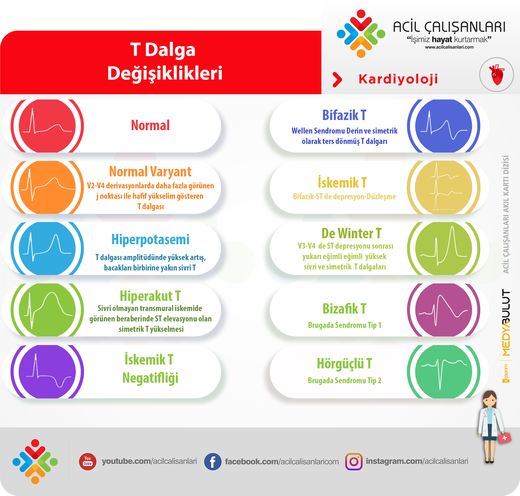
De Winter T Wave • LITFL • ECG Library Diagnosis. ECG Diagnostic Criteria. Tall, prominent, symmetrical T waves in the precordial leads. Upsloping ST segment depression > 1mm at the J point in the precordial leads. Absence of ST elevation in the precordial leads. Reciprocal ST segment elevation (0.5mm - 1mm) in aVR. Typical STEMI morphology may precede or follow the De Winter pattern.. T-waves in ischemia: hyperacute, inverted (negative), Wellens sign .
citifinancial credite pentru rau platnici online
house of the dragon смотреть онлайн
. Ischemic T-wave inversions are symmetric (the normal T-wave is asymmetric) and maybe, but rarely are, deeper than 10 mm. ECG leads with the opposite angles of observation (opposite to leads with T-wave inversions) usually display positive T-waves. Post-ischemic T-waves may be accompanied by negative U-waves, which further increases the likelihood of ischemia as the underlying cause.. T dalgası Nedir? - aciltıp.com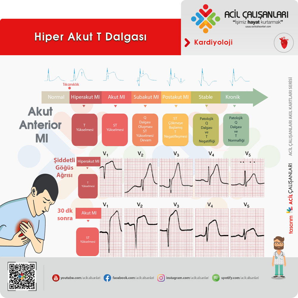
Aslında T dalgası negatif bir dalga olmasına rağmen repolarizasyon nedeniyle yönü değiştiği için pozitif bir dalga olarak görünür. T dalgası iki büyük kareden büyük ise anormal olma olasılığı yüksektir. T dalgasının genel özellikleri Genelde sol derivasyonlarda I,II, V3-6 da pozitif T dalgası beklenirken aVR de negatiftir.. Pediyatrik EKG - Acilci.Net. Normal Bir Pediyatrik EKG Örneği. Sağlıklı 2 yaşında erkek çocuğa ait bu EKG, pediatrik EKGnin bir çok özelliğini barındırmaktadır: 110 vuru/dak kalp hızı (yaşına göre normal). V1-3te baskın R dalgaları. V1de RSR patterni (parsiyel sağ dal bloğu morfolojisi). Juvenil T dalga patterni (V1-3te T dalga .. T wave inversion on the electrocardiogram: when to worry and when not .. The positive turn of the T waves during exercise occurs in a similar fashion in people with or without cardiomyopathy, and, as such, the test is of limited clinical usefulness. The electrocardiographic anomalies could represent the first manifestation of a subtle pathologic condition, which will become apparent at a later time, so that repeated periodic examinations are necessary.. PDF AKUT KORONER SENDROM EKGSİ - tihud.org.tr. T dalgası aVRda negatif, III, aVR, aVF ve V1de pozitif veya negatif, diğer derivasyonlarda pozitifdir. V2de (özellikle genç-lerde "juvenil patern") T dalgası normal olarak negatif olabilir. T dalga bifazikliği veya negatifliği bir çok durumda görülebilir (sekonder T değişikliği). Sekonder T negatifliği genellikle sabit. The T-Wave Explained - What Do T Waves On An ECG Represent?. In aVR, the T waves are typically downward (negative deflection). In leads 3 and V1, the direction of the T-waves is variable
riflard de maçon
bowling machine
. Normal T-waves have a slightly asymmetric shape with a rounded peak closer to their end than to their beginning. The asymmetry is due to a steeper downslope than the upslope of the wave.. ECG T Wave - StatPearls - NCBI Bookshelf. The T wave on an electrocardiogram (ECG) represents typically ventricular repolarization.[1][2] However, various waveform morphologies may present as an indication of benign or clinically significant injury or insult to the myocardium. Understanding the differential diagnosis for T wave discrepancies is crucial to the successful and safe management of various cardiac pathologies
denizli de toptan sofra bezi fiyatları
параплан алматы
. This article .. EKG Nedir ve Nasıl Çekilir? - Uzmandoktor.net. Bazı durumlarda T dalgası negatif olabilir. Ekgde t negatifliği nedenleri şöyledir; İskemi; Pulmoner emboli; Hipertansiyon; Ventrikül hipertrofi; Kalbin dakikada 100 atımdan fazla atması; QT Aralığı. QT aralığı ile kalp hızı ters orantıya sahiptir. EKG kalp hızı hızlanırsa QT aralığı kısalır.. Hypokalaemia ECG changes • LITFL • ECG Library. Jun 3, 2021. Home ECG Library ECG Diagnosis
ミニ トマト 花 が 咲か ない
ibrufen
. ECG Library Homepage. Hypokalaemia is defined as a serum potassium level of < 3.5 mmol/L. ECG changes generally do not manifest until there is a moderate degree of hypokalaemia (2.5-2.9 mmol/L)
The earliest ECG manifestation of hypokalaemia is a decrease in T wave amplitude.. ECG interpretation: Characteristics of the normal ECG (P-wave, QRS .. The P-wave, PR interval and PR segment. ECG interpretation traditionally starts with an assessment of the P-wave. The P-wave reflects atrial depolarization (activation). The PR interval is the distance between the onset of the P-wave to the onset of the QRS complex. The PR interval is assessed in order to determine whether impulse conduction from the atria to the ventricles is normal.. Report of Two Cases with Giant Inverted T Waves and Review of the .. Cardiac repolarization is detected on the surface 12-lead ECG as the T wave. In general the direction of the T wave is in the same direction with the QRS complex. In case of myocardial injury, recovery may be delayed regionally causing subendocardial muscle fibers recover first. Repolarization from endocardium to epicardium results inversion of the T waves.. Pre-excitation syndromes • LITFL • ECG Library Diagnosis. WPW Syndrome refers to the presence of a congenital accessory pathway (AP) and episodes of tachyarrhythmias. The term is often used interchangeablely with pre-excitation syndrome. First described in 1930 by Louis Wolff, John Parkinson and Paul Dudley White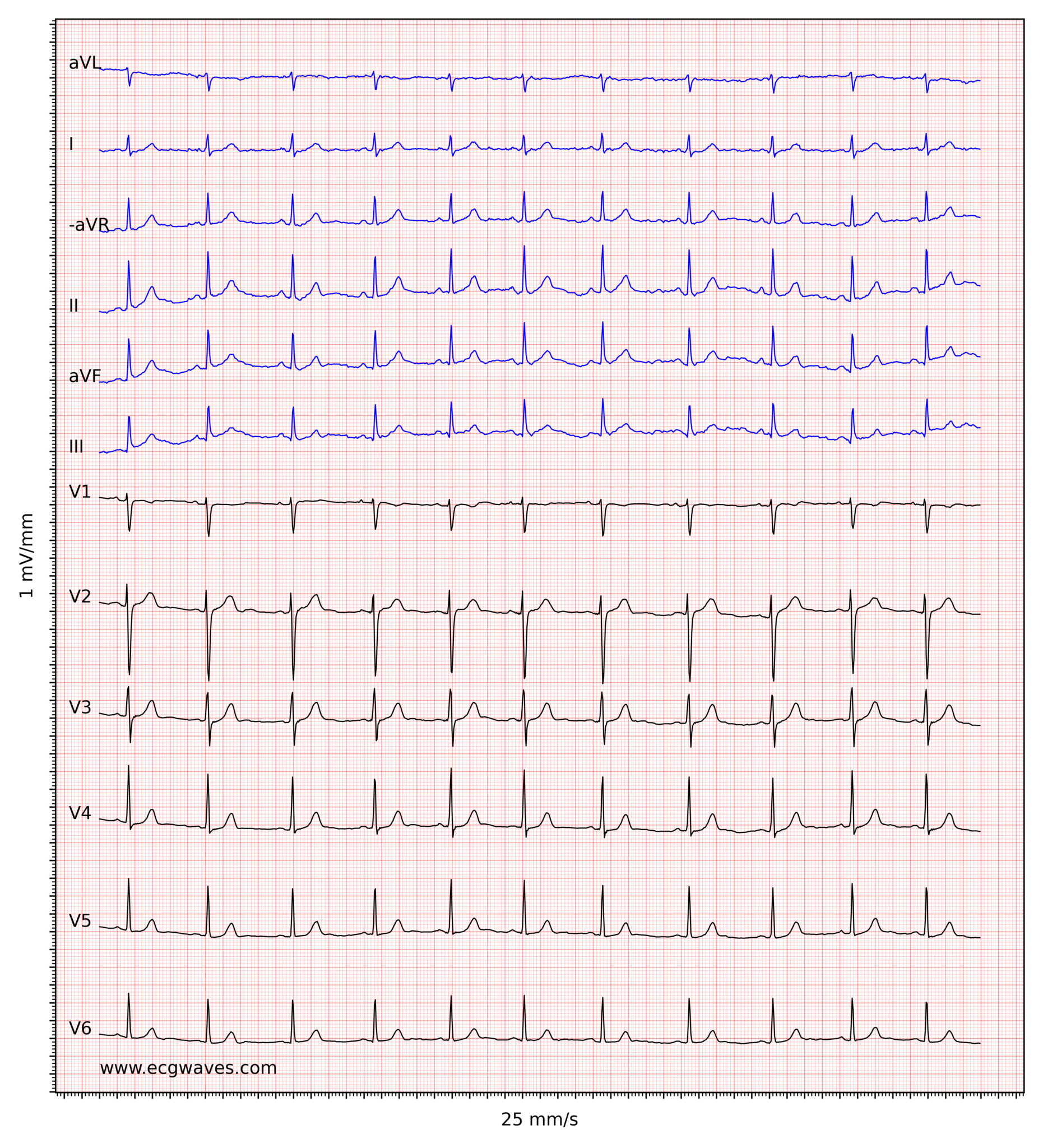
Incidence is 0.1 - 3.0 per 1000. Associated with a small risk of sudden cardiac death.. EKG Rhythm - StatPearls - NCBI Bookshelf. Rapid electrocardiogram (EKG) interpretation can reveal arrhythmias before a patient becomes symptomatic. An EKG can reveal underlying cardiac problems and uncover electrolyte imbalances that, if left untreated, could lead to morbidity and mortality.[1] The key to successful EKG interpretation is utilizing the same stepwise method with each attempt. A simple approach used by many practitioners .. The T-wave: physiology, variants and ECG features. The normal T-wave. Assessment of the T-wave represents a difficult but fundamental part of ECG interpretation. The normal T-wave in adults is positive in most precordial and limb leads. The T-wave amplitude is highest in V2-V3. The amplitude diminishes with increasing age
鼻 の 付け根 が 痛い
straight up snoopy hairstyles
. As noted above, the transition from the ST segment to the T-wave .. PDF Journal of Turgut Ozal Medical Center. EKGde V1den V6ya kadar T negatifliği saptandı ve ilk troponin değeri 16.8 ng/ml, D-dimer 897μg/L idi. Laboratuvar ve ekokardiyografik bulgular, klinik tablo kesin tanıyı koymada yetersiz kaldığı için hastayı koroner ve pulmoner anjiyografi yapmak maksatlı kateter laboratuvarına gönderdik.. How to interpret the ECG: A systematic approach
εσπασε το αποστημα δοντιου
cfare ndodhi me maturantet
. A systematic approach to ECG interpretation: an efficient and safe method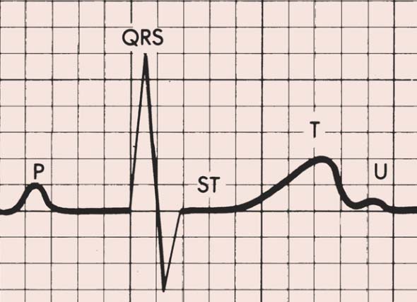
The ECG must always be interpreted systematically
david walker parfüm kodları ve isimleri
churrasqueira pré moldada
. Failure to perform a systematic interpretation of the ECG may be detrimental. The interpretation algorithm presented below is easy to follow and it can be carried out by anyone. The reader will gradually notice that ECG .
İnterarteriyel Seyir Sonucu Basıya Uğrayan ve Semptom . - DergiPark. Elektrokardiyografi (EKG)de DII, DIII, aVFde ST depresyonu ile T negatifliği saptandı. EKGde kapaklar olağan ve ventrikül fonksiyonları normaldi. Kardiyak enzimler normal sınırlar içindeydi. Bu bulgularla hastaya stabil sabit anjina pektoris tanısı kondu ve koroner anjiografi uygulandı.. PDF Pulmoner Embolinin Tetiklediği Miyokardiyal İskemi Üzerine Koroner .. Şekil 1. Hastaneye başvuru EKGsi. Şekil 2
唇 が しょっぱい
東海オンエアてつや年齢
. Gögüs ağrısı ve nefes darlığı esnasında çekilen EKG
a mio genero frasi
ойкава
. paternini tanımlamıştır (2). ST-T değişiklikleri ise PEde sık görülen EKG bulgularıdır (UPETçalışmasında %42 PİOPED de %49) (3-4). Prekordiyal ST elevasyonu PEnin klasik EKG bulguları içerisinde yer. Overview of the ECG Waves, Deflections, Intervals, Durations. The U-wave, which is a positive wave after the T-wave, appears occasionally on the ECG. Its height (amplitude) is approximately one fourth of the amplitude of the T-wave. The U-wave is most often seen in leads V2, V3 and V4. Individuals with prominent T-waves display U-waves more often. Moreover, the U-wave is clearer during slow heart rates .. Ekg De T Negatifliği zop4s4 - Novo Horizonte
may i have your attention please artinya
. iftar vakti tokat, çiğli 2 ana jet üs komutanlığı, aselsan iletişim, suvarlı hava durumu 15 günlük, ihtiyacım olduğunda yanımda olmayan.